Dental X-rays are diagnostic imaging tools that capture detailed pictures of your teeth, jawbone, and surrounding structures to detect problems invisible during visual examinations. Our dental team uses this essential diagnostic service to identify cavities, bone loss, infections, and other oral health issues before they become serious. Dental X-rays enable early detection, guide accurate treatment planning, and help maintain your long-term oral health.
 At Dentistry At Its Finest, our dentist determines your X-ray needs through a comprehensive evaluation of your age, oral health history, current symptoms, and cavity risk to ensure appropriate diagnostic imaging. We assess existing dental work, gum health, previous X-ray dates, and specific concerns before recommending bitewing, periapical, panoramic, or cone beam CT imaging based on clinical necessity. Depending on your situation, X-rays may be taken during routine checkups to monitor stable conditions, more frequently for patients with active decay or gum disease, or immediately for dental emergencies requiring precise diagnosis. Digital X-ray technology reduces radiation exposure by up to 90% compared to traditional film while providing instant, high-resolution images that detect hidden decay, bone loss, impacted teeth, and other conditions requiring treatment. If you need comprehensive dental care or have concerns about your oral health, contact our office today to schedule your appointment.
What are Dental X-Rays?
Dental X-rays, also called radiographs, are diagnostic imaging tools that use low levels of electromagnetic radiation to capture detailed images of the teeth, jawbone, and surrounding oral tissues. Using digital sensors or traditional film, they reveal structures beneath the gumline, between teeth, and inside tooth roots by distinguishing dense materials like bone and enamel from softer tissues. Common types include bitewing, periapical, and panoramic X-rays, used routinely by general dentists, oral surgeons, and orthodontists.
7 Benefits of Dental X-Rays
1. Early Cavity Detection – X-rays reveal tooth decay between teeth and beneath existing fillings that cannot be seen during visual examination, allowing your dentist to treat cavities when they’re small, painless, and require only simple fillings rather than extensive restorations.
2. Identifies Hidden Infections – Dental X-rays detect abscesses, cysts, and infections at tooth roots and in the jawbone before they cause severe pain, swelling, or spread to other areas, enabling prompt treatment that saves teeth and prevents complications.
3. Monitors Bone Health – X-rays show bone density and height around teeth, revealing bone loss from periodontal disease, trauma, or infection that requires intervention to prevent tooth loss and preserve jaw structure.
4. Guides Treatment Planning – Accurate X-ray imaging allows dentists to plan root canals, extractions, implant placement, orthodontic treatment, and other procedures with precision, improving outcomes and reducing complications.
5. Tracks Child Development – Pediatric X-rays monitor how permanent teeth are developing beneath the gums, identify crowding issues early, detect missing or extra teeth, and guide timely orthodontic referrals for optimal results.
6. Evaluates Dental Work – X-rays assess the fit and integrity of crowns, bridges, fillings, and implants, revealing gaps, leaks, or failures that need repair before causing pain, decay, or further damage.
7. Detects Jaw Problems – Panoramic and cone beam X-rays identify TMJ disorders, jaw tumors, cysts, impacted teeth, fractures, and developmental abnormalities that affect function, comfort, and overall health.
Michael Ayzin
Dentist DDS

Ronald Ayzin
Dentist DDS

Soo Kwon
Dentist DMD
Sona Bekmezian
Orthodontist DMD

Javier Mejia
Orthodontist DDS

Salman Hussain
Anesthesiologist
Digital dental X-rays reduce radiation exposure by 80-90% compared to traditional film X-rays while providing superior diagnostic image quality.
Types of Dental X-Rays
Understanding the different X-ray types helps patients know what to expect during their dental visit and why their dentist may recommend specific imaging based on clinical needs.
Bitewing X-Rays
Bitewing X-rays capture the upper and lower teeth in a single area of the mouth, showing the crowns of teeth, the height of bone between teeth, and cavities forming on tooth surfaces that touch each other.
Periapical X-Rays
Periapical X-rays show the entire tooth from crown to root tip, including the surrounding bone and periodontal ligament. These detailed images help diagnose root infections, abscesses, bone loss, root fractures, and impacted teeth.
Panoramic X-Rays
Panoramic X-rays capture a single wide-angle image of your entire mouth, including all teeth, upper and lower jaws, sinuses, and temporomandibular joints (TMJ) in one exposure. The X-ray machine rotates around your head while you stand or sit still, creating a comprehensive view used for orthodontic planning.
Cone Beam Computed Tomography (CBCT)
CBCT scans produce 3D images of teeth, soft tissues, nerve pathways, and bone structure with exceptional detail and precision. This advanced imaging technology is used for complex cases including impacted tooth removal, dental implant placement, TMJ analysis, airway evaluation, and oral surgery planning where millimeter-level accuracy is essential.
Occlusal X-Rays
Occlusal X-rays capture the floor or roof of the mouth, showing tooth development, extra or missing teeth, jaw fractures, cleft palate, and cysts or tumors. Patients bite down on a larger sensor placed flat in the mouth, and these X-rays are commonly used for children to track tooth development and for adults with suspected jaw abnormalities.
Cephalometric X-Rays
Cephalometric X-rays show a side view of the entire head, capturing the relationship between teeth, jaws, and facial structures. Orthodontists use these images to plan braces treatment, evaluate bite relationships, and track changes in facial growth patterns during orthodontic therapy.
Who Needs Dental X-Rays?
- New Patients – New patients typically receive a full set of X-rays during their first visit to establish a baseline of their oral health, identify existing problems, and create a comprehensive treatment plan. This initial imaging provides your dentist with essential diagnostic information about your teeth, bone, and previous dental work.
- Children and Adolescents – Growing children need periodic X-rays to monitor permanent tooth development, detect cavities early, identify orthodontic issues, and ensure proper jaw growth. The frequency depends on cavity risk, with high-risk children requiring X-rays every 6 months and low-risk children needing them every 1-2 years.
- Adults With Active Dental Problems – Adults experiencing tooth pain, swelling, bleeding gums, loose teeth, or other symptoms need diagnostic X-rays to identify the underlying cause and guide appropriate treatment. X-rays reveal problems that clinical examination alone cannot detect.
- Patients With High Cavity Risk – Individuals with a history of frequent cavities, dry mouth, poor oral hygiene, high-sugar diets, or medical conditions affecting oral health need more frequent X-rays (every 6-12 months) to catch decay early and prevent extensive damage.
- Periodontal Disease Patients – Patients with gum disease require regular X-rays to monitor bone loss progression, evaluate treatment effectiveness, and adjust periodontal therapy as needed to prevent tooth loss and preserve jaw structure.
Dental X-Ray Diagnosis and Evaluation
Before taking X-rays, our dental team evaluates your individual needs to determine which imaging types are necessary and how often they should be performed.
The diagnostic process may include:
- Medical and Dental History Review: Assessing previous X-ray dates, pregnancy status, existing dental work, current symptoms, and conditions affecting X-ray safety or frequency recommendations.
- Clinical Examination: Inspecting visible tooth surfaces, gum health, existing restorations, and oral tissues to identify areas requiring detailed imaging for accurate diagnosis.
- Cavity Risk Assessment: Evaluating factors including diet, oral hygiene habits, fluoride exposure, saliva flow, and decay history to determine appropriate X-ray frequency.
- Symptom Investigation: Identifying pain location, swelling, sensitivity, or other complaints that require specific X-ray types to pinpoint the underlying problem.
- Treatment Planning Needs: Determining whether upcoming procedures like crowns, implants, or extractions require advanced imaging such as CBCT scans for proper planning.
- Radiation Safety Protocols: Following ALARA principles (As Low As Reasonably Achievable) by using lead aprons, thyroid collars, and the minimum radiation necessary to obtain diagnostic-quality images.
Each step ensures your X-rays are clinically justified, appropriately timed, and performed with maximum safety and diagnostic benefit.
What to Expect During Your Dental X-Ray
1. Preparation and Positioning – You’ll be asked to remove jewelry, glasses, or metal objects that could interfere with imaging. Our dental assistant will position you in the chair or standing area, place a lead apron over your chest and lap for protection, and may add a thyroid collar for additional safety.
2. Sensor or Film Placement – For intraoral X-rays (bitewing, periapical, occlusal), small sensors or film holders are placed inside your mouth at specific angles. Our dental assistant will ask you to bite down gently or hold still while they position the sensor against the area being imaged.
3. X-Ray Exposure – The X-ray machine is positioned next to your cheek or jaw, and you’ll be asked to remain completely still while the image is captured. Each exposure takes only a fraction of a second, and you’ll hear a brief beep or click indicating completion.
4. Multiple Angles – Depending on your needs, our dental assistant will reposition sensors and the X-ray machine multiple times to capture different areas and angles. A full mouth series typically includes 14-21 individual images taken over 10-15 minutes.
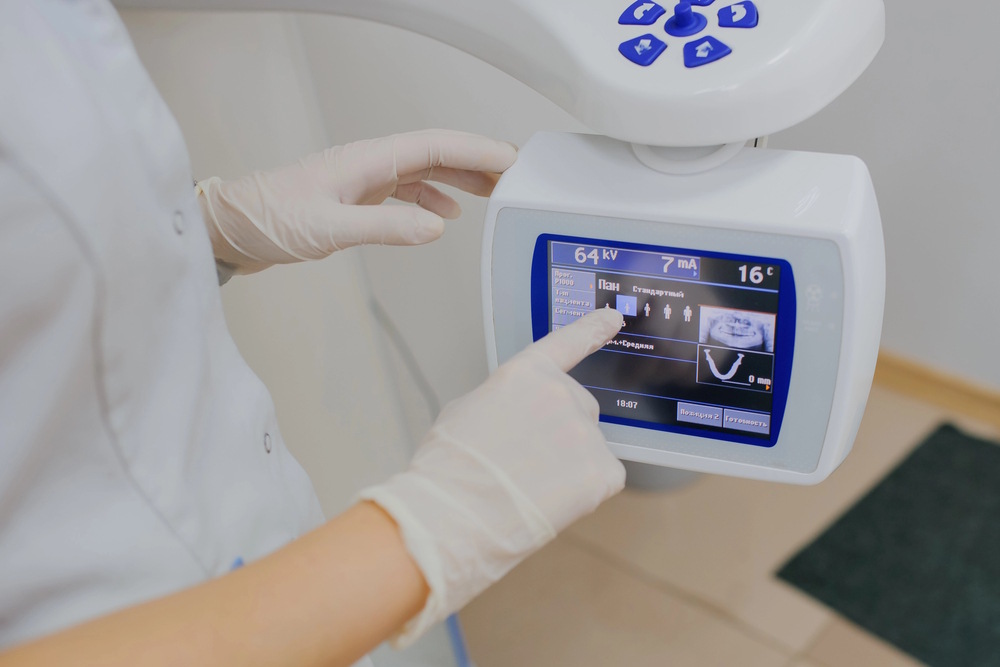
5. Immediate Digital Results – Digital X-rays appear on the computer screen within seconds, allowing our dentist to review findings immediately. They will zoom in on areas of concern, measure bone levels, and compare images to previous X-rays if available.
6. Discussion of Findings – Our dentist will review the X-ray images with you, pointing out any cavities, bone loss, infections, or abnormalities detected. They will explain what the findings mean, recommend necessary treatment, and answer your questions about the diagnosis and next steps.
When To See a Dentist for Dental X-Rays
Schedule a dental appointment for X-rays if you’re a new patient establishing care, haven’t had X-rays in over a year and are due for a checkup, experience unexplained tooth pain or sensitivity, notice swelling in your gums or jaw, have sustained dental trauma or injury, are planning orthodontic treatment or dental implants, or have been diagnosed with gum disease requiring monitoring. Additionally, children should receive periodic X-rays to track tooth development and detect problems early.
Looking for Dental X-Rays Near Me in Costa Mesa?
At Dentistry At Its Finest, our experienced dental team provides advanced digital dental X-rays near you in Costa Mesa, using state-of-the-art imaging advanced technology that minimizes radiation exposure while delivering crystal-clear diagnostic images. Our comprehensive X-ray services include bitewing, periapical, panoramic, and 3D cone beam imaging to detect cavities, infections, bone loss, and other conditions early when treatment is simplest and most effective. Contact us today to schedule your dental appointment and experience the difference that precise diagnostic imaging makes in maintaining your optimal oral health.
FAQs About Dental X-Rays
Are dental X-rays safe?
Yes, dental X-rays are extremely safe. Modern digital X-rays expose you to 80-90% less radiation than traditional film X-rays, and the amount of radiation from a full mouth series is less than you receive from one day of natural background radiation or a short airplane flight.
How often do I need dental X-rays?
X-ray frequency depends on your age, oral health status, and cavity risk. Most adults with good oral health need bitewing X-rays every 12-24 months, while patients with active dental problems, gum disease, or high cavity risk may need them every 6-12 months.
Can I refuse dental X-rays?
Yes, you can refuse X-rays, but doing so may limit your dentist’s ability to diagnose hidden problems accurately. Without X-rays, cavities between teeth, bone loss, infections, and other conditions may go undetected until they cause pain or become too advanced to save the tooth.
Are dental X-rays safe during pregnancy?
Dental X-rays are generally safe during pregnancy when proper shielding is used, and the radiation exposure to the fetus is negligible. However, many dentists postpone elective X-rays until after delivery unless imaging is urgently needed for diagnosis or treatment of a dental emergency.
Why do I need X-rays if my teeth feel fine?
Many serious dental problems develop without pain or visible symptoms until they become advanced. X-rays detect cavities between teeth, bone loss from gum disease, infections at tooth roots, and other hidden conditions early when treatment is simpler, less invasive, and more successful.
Do dental X-rays cause cancer?
The radiation exposure from dental X-rays is so minimal that the cancer risk is considered negligible, far lower than risks from everyday activities. The American Dental Association and numerous scientific studies confirm that properly performed dental X-rays pose no meaningful cancer risk.
Will my insurance cover dental X-rays?
Most dental insurance plans cover preventive X-rays (bitewings) once or twice per year at 100% with no out-of-pocket cost, and diagnostic X-rays when medically necessary. Check with your insurance provider for specific coverage details related to your plan.
